MEET OUR CODE_N CONTEST FINALISTS 2018: OXIPIT from Lithuania
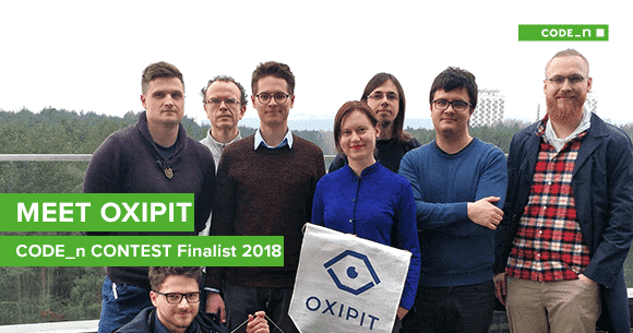 AI applications extend to a myriad of industries, including healthcare. The capability to radically transform lives with pioneering technologies is one of the immeasurable benefits of AI. One such example is AI for medical image diagnosis. Headquartered in Vilnius, OXIPIT, a CODE_n CONTEST finalist is an AI-powered medical imaging startup focusing on improving the accuracy of radiological analysis for many kinds of pathologies. In this interview, Jogundas Armaitis, COO of OXIPIT, lets us know how their solution helps to save time and facilitate better diagnostic outcomes.
Aishah: What is OXIPIT all about?
Jogundas: OXIPIT is all about accelerating clinical adoption of the recent advances in AI. Basically, our software takes in an image and describes the relevant findings in plain text. In particular, we are applying AI (computer vision and deep learning) to radiology. A radiologist is a highly specialized doctor who analyzes medical images and describes them in text. These reports are then read by other clinicians, who in turn treat the patient. Radiology is a vast field, covering different imaging methods (X-ray, CT, MRI etc.) that are applied to different parts of the human body. We are specialized in chest X-rays, which are the most common radiological examination and are still increasing in number. The average person in the developed world gets a chest X-ray every four years. Our claim to fame is our stellar data science team. We’ve won multiple international competitions, including the Intel & MobileODT Cervical Cancer Screening competition. Our co-founder Darius is currently ranked fourth out of almost 100,000 data scientists on Kaggle.
Aishah: How did you come up with the idea?
Jogundas: It all started back in April 2017, when some of the future OXIPIT founders mentored several teams at AI camp, which is a micro-conference and hackathon in Vilnius. One of the teams there applied AI to medical images, and they won the hackathon. The winning team leader Naglis, a radiologist, approached the rest of us after the event and proposed that we start a medical imaging company together. We were skeptical at first, since obtaining enough medical data to design an AI product looked like a daunting proposition. However, after extensive discussions with numerous doctors throughout the summer of 2017, we had a list of medical imaging projects which were both clinically relevant and also had data available. After further analysis of these clinical projects, we chose the one with the highest impact.
Aishah: What are you trying to solve?
Jogundas: We are trying to solve issues surrounding chest X-rays, the most common radiological examination. The hardware usually exists to acquire the right images, but often there’s a lack of human resources to interpret them. Chest X-rays offer numerous advantages (low radiation, mature hardware, standardized images, cheap to perform), but there are also some challenges, the most severe of which is the shortage of highly qualified radiologists to analyze the images. We’re addressing this challenge by creating AI tools for radiologists. Our ultimate aim is to add value for patients (more accurate diagnosis), physicians (fewer routine tasks), and medical providers (more productive radiologists).
Aishah: Is your solution ready to be used in hospitals? What feedback have you received from radiologists so far?
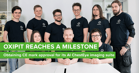
Jogundas: We receive continual feedback from the dozens of radiologists trying out the OXIPIT solution. The feedback we’re getting is extremely positive. It’s important to add that our software is not yet available for clinical use in the European Union because we’re still in the process of certifying it. In addition to delivering reliability and accuracy, we believe that the product must be convenient to use. In our case, it’s important not to alter the current radiological workflow too much. To that end, we’ve been working on integrating our solution into other software such as radiological image (DICOM) viewers or picture archiving and communication systems (PACS). We’re currently at the point where radiologists can use our solution without changing their current workflows. Besides qualitative feedback, we have also run a controlled trial to measure how our product compares to productivity tools already being routinely used by radiologists today, such as templates. The results of the trial were great: It turns out that our solution offers 30% time savings and cuts the number of reporting error by up to two thirds in comparison to regular workflows.
Aishah: Thanks so much for the interview Jogundas and all the best to the OXIPIT team!
Meet OXIPIT at the new.New Festival 2018 this fall, in Stuttgart!

By loading the video, you agree to YouTube’s privacy policy.
Learn more

Write a comment